This page contains specialized information intended exclusively for licensed medical professionals. By continuing, you confirm that you are a physician or medical service provider.
Precise Knee Cartilage Repair
For patients with focal cartilage defects of the knee, TETEC offers two autologous, cell-based approaches: Injectable MACT (Matrix-Associated Autologous Chondrocyte Transplantation) and MACT. Both use the patient's own cultured chondrocytes to support cartilage repair. Continue exploring to learn more about each procedure—and where they fit within the current clinical landscape.
.avif)
Focal Knee Cartilage Lesions
Each year in Germany, tens of thousands of knee-cartilage procedures are performed. If left untreated, focal chondral defects may enlarge and, in some patients, contribute to degenerative joint changes consistent with osteoarthritis [1, 2, 3].
Typical Aetiologies
Acute trauma (sports injuries, high-impact twists)
Repetitive Overload
Osteochondritis dissecans (OCD)
Malalignment or chronic instability
Why Cartilage Heals Poorly
Avascular cartilage limits nutrients and repair cells
Low cell turnover slows matrix regeneration
Dense extracellular matrix blocks reparative cell migration
Constant joint loading disrupts repair and drives breakdown
Compare Leading Techniques
Traditional cartilage repair techniques—such as Bone Marrow Stimulation (BMS), Matrix-Augmented BMS, Osteochondral Autograft Transfer System (OATS), and others shown below are limited by lesion size, surgical complexity, and whether inpatient treatment is required.
.jpeg)
Injectable MACT vs. MACT
Explore Injectable MACT and MACT in detail.
Knee Cartilage Repair With Cells
TETEC develops two personalised treatments for knee cartilage repair, both using the patient's own cells: Injectable MACT and scaffold-based MACT.
Injectable MACT Procedure
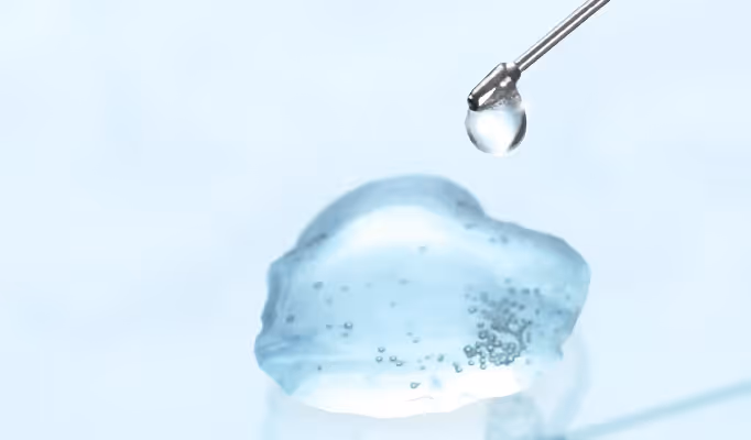
Injectable MACT is a cell-based cartilage-repair procedure in which a patient's own cartilage cells are combined with a hydrogel and implanted into a focal knee cartilage defect [6]. In clinical studies and earlier publications, this approach is also described as hydrogel-based ACI or injectable MACI.
- Injectable hydrogel containing the patient's cartilage cells
- Implanted arthroscopically through small incisions
- Available as outpatient or inpatient care, as appropriate
MACT Procedure
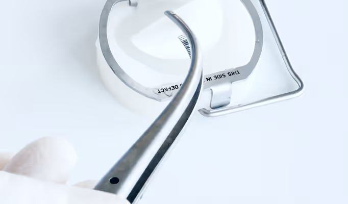
In MACT, the patient's own cells are placed on a collagen scaffold and implanted for the treatment of focal knee cartilage defects [5].
- Patch-like scaffold seeded with the patient's cartilage cells
- Implanted through a standard surgical incision
- Available as outpatient or inpatient care, as appropriate
Stay Updated on TETEC
Discover TETEC news and stay informed about upcoming conferences.
1. Ding C, Cicuttini F, Scott F, Boon C, Jones G. Association of prevalent and incident knee cartilage defects with loss of tibial and patellar cartilage: a longitudinal study. Arthritis Rheum. 2005;52(12):3918–3927.
2. Cicuttini F, Ding C, Wluka A, Davis S, Ebeling PR, Jones G. Association of cartilage defects with loss of knee cartilage in healthy, middle-aged adults: a prospective study. Arthritis Rheum. 2005;52(7):2033–2039.
3. Houck DA, Kraeutler MJ, Belk JW, Frank RM, McCarty EC, Bravman JT. Do focal chondral defects of the knee increase the risk for progression to osteoarthritis? A review of the literature. Orthop J Sports Med. 2018;6(10):2325967118801931. doi:10.1177/2325967118801931.
4. Niemeyer P, Albrecht D, Aurich M, Becher C, Behrens P, Bichmann P, et al. Empfehlungen der AG Klinische Geweberegeneration zur Behandlung von Knorpelschäden am Kniegelenk. Z Orthop Unfall. 2023;161(1):57–64. doi:10.1055/a-1663-6807.
5. TETEC AG. Fachinformation NOVOCART 3D. Reutlingen (DE): TETEC AG; 2023. Stand der Information: 11/2023. PEI.A.11511.01.1.
6. TETEC AG. Fachinformation NOVOCART Inject. Reutlingen (DE): TETEC AG; 2022. Stand der Information: 12/2022. PEI.A.11763.01.1.
7. Niemeyer P, Hanus M, Belickas J, László T, Gudas R, Fiodorovas M, et al. Treatment of large cartilage defects in the knee by hydrogel-based autologous chondrocyte implantation: two-year results of a prospective, multicenter, single-arm phase III trial. Cartilage. 2022;13(1):19476035221085146. doi:10.1177/19476035221085146.
8. Paul-Ehrlich-Institut. Physician training guide for NOVOCART 3D. Langen (DE): Paul-Ehrlich-Institut; 2025. Available from: https://www.pei.de/SharedDocs/schulungsmaterial/Novocart-3D-Schulungsmaterial-Aerzte_Version-2_Leitfaden.pdf
9. Schlumberger M, Schuster P, Bülow HJ, Mayer P, Eichinger M, Richter J. Arthroscopic autologous chondrocyte implantation in the knee with an in situ crosslinking matrix: minimum 4-year clinical results of 15 cases and 1 histological evaluation. Arch Orthop Trauma Surg. 2019;139(11):1607–1615.
10. Data on File. TETEC AG. Internal registry and reporting of MACT and Injectable MACT procedures performed in Germany. Reutlingen (DE): TETEC AG; 2025.