A Knee Cartilage Treatment Using Your Own Cells
MACT (matrix-associated autologous chondrocyte transplantation) is a form of autologous chondrocyte implantation (ACI) in which the patient's own cartilage cells are placed on a biphasic collagen scaffold to treat damaged areas of the knee. This method, which has been used in routine clinical care for over 20 years, is also described as MACI or M-ACI.

Understanding the Basics

Long-Lasting Outcomes
Sustained clinical outcomes for MACT have been reported up to 10 years after treatment [1, 2].
Established Treatment
Recognised as an option for symptomatic knee cartilage defects larger than 2 cm², where appropriate [3, 4].
Your Own Cells
The MACT procedure seeds a collagen scaffold with your own cartilage cells, which contribute to the formation of repair tissue [5].
How It Works
Once a full-thickness defect has been diagnosed by a specialist, MACT proceeds in four phases.
Attend Your Biopsy
During the diagnostic arthroscopy, small cartilage-bone cylinders are taken from the non-weight-bearing area of the knee.
We Prepare Your Graft
At TETEC, the cartilage cells from this biopsy are isolated, expanded in the laboratory, and seeded onto a 3D collagen scaffold. This forms the basis of the MACT graft.
.avif)
Receive Your Graft
The collagen scaffold is then implanted into the damaged area of the knee during a small surgical procedure.
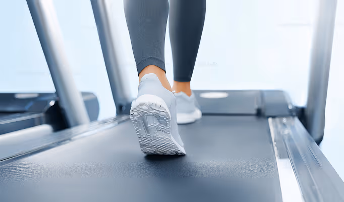
Recovery and Rehabilitation
The typical recovery period involves 48 hours without knee mobilisation; walking aids may be used during recovery. Rehabilitation depends on the defect location, with physiotherapy and MRI at approximately 24 months [6].
When Is MACT Used?
Autologous chondrocyte implantation is generally suitable for the surgical reconstruction of full-thickness cartilage damage in the knee. In addition to lesion size and cartilage condition, other factors play an important role.
The right treatment for knee cartilage damage can depend on factors such as biological age, lifestyle, and body weight. Your specialist will determine whether MACT is suitable for you after a thorough examination. These individual factors must always be assessed by the treating orthopaedic surgeon.

Important Notes
Post-operative care following cartilage cell implantation depends primarily on the anatomical location of the cartilage defect in the knee joint. The cartilage must first fully heal in this position in order to optimally resume its role as a shock absorber for the joint. Gradual muscular stabilisation of the joint is extremely important and can take a great deal of time.
Since every individual responds differently to surgical intervention, the post-treatment programme may vary from patient to patient. As a rule, the joint should be kept still for the first 48 hours after surgery. After that, gentle movement is introduced. Depending on the location of the defect, walking aids are used to avoid loading the knee. Physiotherapy continues in the following weeks. Your doctor will determine the appropriate measures for you after surgery.
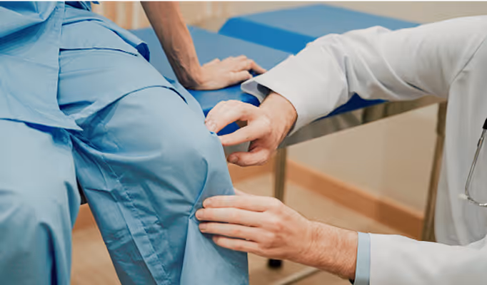
- Medical History
Doctor asks about how the injury happened, symptoms (pain, swelling, locking), physical activity, and prior knee issues. - Physical Examination
Manual tests to assess pain location, joint stability, swelling, range of motion, and signs of mechanical symptoms (e.g. catching, grinding). - Imaging
- X-ray: Rules out bone fractures or arthritis but can’t show cartilage directly.
- MRI: For visualizing cartilage defects, soft tissues, and bone bruising.
- CT or CT-Arthrography: Used when MRI is unclear or unavailable.
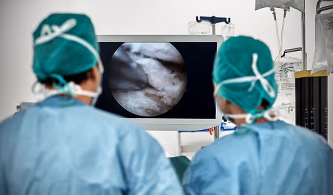
- Arthroscopy (Optional)
Minimally invasive camera-based procedure. Directly inspects cartilage and may be used if imaging is inconclusive or surgery is being planned. - Grading the Damage
Severity is classified by systems like ICRS Grade I–IV, based on how deep and wide the defect is.
Consult your physician or view our treatment centres.

Is This Procedure an Option?
Only a medical examination can confirm whether TETEC solutions are suitable for your knee. Our procedures are not recommended for advanced or extensive osteoarthritis.
1. Weishorn J, Wiegand J, Zietzschmann S, Koch KA, Rehnitz C, Renkawitz T, Walker T, Bangert Y. Factors influencing long-term outcomes after matrix-induced autologous chondrocyte implantation: long-term results at 10 years. Am J Sports Med. 2024;52(11):2782–2791. doi:10.1177/03635465241270152.
2. Weishorn J, Niemeyer P, Walker T, Tsitlakidis S, Renkawitz T, Bangert Y. Favourable clinical outcomes and low revision rate after M-ACI in adolescents with immature cartilage compared to adult controls: results at 10 years. Knee Surg Sports Traumatol Arthrosc. 2025;33(1):167–176. doi:10.1002/ksa.12359.
3. National Institute for Health and Care Excellence (NICE). Autologous chondrocyte implantation for treating symptomatic articular cartilage defects of the knee. Technology appraisal guidance TA477. London: NICE; 4 October 2017.
4. Niemeyer P, Albrecht D, Aurich M, et al. Empfehlungen der AG Klinische Geweberegeneration zur Behandlung von Knorpelschäden am Kniegelenk. Z Orthop Unfall. 2023;161(1):57–64. doi:10.1055/a-1663-6807.
5. Eichinger M, Henninger B, Petry B, Schuster P, Herbst E, Wagner M, Rosenberger R, Mayr R. Treatment of cartilage defects in the patellofemoral joint with matrix-associated autologous chondrocyte implantation effectively improves pain, function, and radiological outcomes after 5–7 years. Arch Orthop Trauma Surg. 2024;144(4):1655–1665. doi:10.1007/s00402-023-05179-0.
6. Data on file. TETEC AG. Internal registry and reporting of Injectable MACT and MACT procedures performed in Germany. Reutlingen (DE): TETEC AG; 2025.