This page contains specialised information intended exclusively for licensed medical professionals. By continuing, you confirm that you are a physician or medical care provider.
MACT: Matrix-Associated Autologous Chondrocyte Transplantation
MACT is a form of Autologous Chondrocyte Implantation (ACI) that uses the patient’s own chondrocytes on a collagen scaffold to help repair focal knee cartilage defects. Once implanted, the cells support regeneration of cartilage-like repair tissue.
Indicated for Focal Knee Lesions
For symptomatic full-thickness cartilage defects, ICRS grade III–IV, 2–10 cm² [1, 2].
Delivered Through Mini-Arthrotomy
Implanted via mini-arthrotomy, fixed with sutures [1]. Established Collagen-Scaffold Technology
Established Collagen-Scaffold Technology
A biphasic collagen scaffold keeps cells localised within the defect [1].
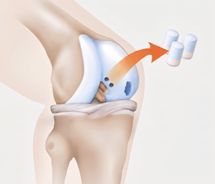
Understanding the Process
In MACT, autologous chondrocytes are implanted into the defect. Over time, these cells support regeneration of cartilage with durable biomechanical properties. The procedure involves biopsy, processing, and implantation back into the defect area via mini-arthrotomy.
Cartilage Biopsy
During diagnostic arthroscopy, small cartilage-bone cylinders are taken from a non-weight-bearing part of the joint.
.jpg)
Cell Cultivation
At TETEC, the chondrocytes from this biopsy are isolated, expanded in vitro, and applied to a three-dimensional biphasic collagen scaffold.
.jpg)
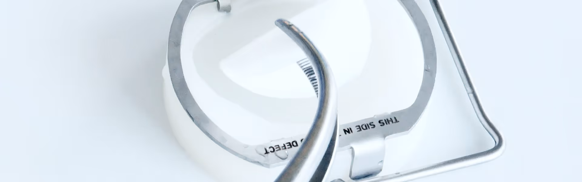
MACT Procedure
The cell-seeded collagen scaffold can then be transplanted into the cartilage defect in a mini-arthrotomy procedure.
.png)
Recovery and Rehabilitation
Standard rehabilitation is defect-location based and DGOU-oriented. It requires 48 hours without knee mobilisation before controlled joint movement can be initiated [1].
For load-bearing femoral condyle defects,partial weight-bearing is limited to a max of 20 kg during weeks 1–6.Two forearm crutches are used until full weight-bearing is achieved [1].
For patellar or retropatellar defects, full weight bearing is generally permitted, with a ROM brace and flexion limits determined by defect size and location [1].
Distinct Properties of MACT
MACT has distinct clinical properties and is supported by published clinical experience, including mid- to long-term follow-up (see Clinical Outcomes).
01
Autologous chondrocytes (no donor tissue; low immunogenicity)
04
Typically performed as an inpatient procedure
02
Established collagen scaffold supports cartilage regeneration
05
Published mid- to long-term outcomes reported in multiple studies
03
Defect coverage for ~2–10 cm²
Indications
When to Use Our Products
MACT is promising in the case of full-thickness and symptomatic cartilage defects surrounded by healthy cartilage. Patients with advanced osteoarthritis are no longer candidates for biological cartilage reconstruction and require a different treatment, such as an artificial joint replacement.
Choosing the Right Therapy
As with many injuries, illnesses and therapies, individual factors play an important role in choosing MACT. In addition to the cartilage’s properties, determining the most suitable therapy will depend on a range of criteria, including the following:
Previous treatments
Weight
Biological age
Patient’s physical and sporting activities
Comorbidities and risk factors (e.g., smoking, metabolic disease)

Clinical Outcomes
Clinical studies report favourable mid-term (5–7 years) outcomes after collagen-scaffold MACT, with additional cohorts reporting durable results and low revision rates at long-term follow-up (10 years) [3, 4, 5]. Patients with localized degenerative cartilage damage may also benefit [1], with prospective series reporting encouraging outcomes at follow-up to 15 years [6].

Why Choose MACT?
Contact TETEC
Regulatory / Product Information
1. TETEC AG. Fachinformation NOVOCART 3D. Reutlingen (DE): TETEC AG; 2023. Stand der Information: 11/2023. PEI.A.11511.01.1.
2. Niemeyer P, Albrecht D, Aurich M, Becher C, Behrens P, Bichmann P, et al. Empfehlungen der AG Klinische Geweberegeneration zur Behandlung von Knorpelschäden am Kniegelenk. Z Orthop Unfall. 2023;161(1):57–64. doi:10.1055/a-1663-6807.
3. Eichinger M, Henninger B, Petry B, Schuster P, Herbst E, Wagner M, Rosenberger R, Mayr R. Treatment of cartilage defects in the patellofemoral joint with matrix-associated autologous chondrocyte implantation effectively improves pain, function, and radiological outcomes after 5–7 years. Arch Orthop Trauma Surg. 2024;144(4):1655–1665. doi:10.1007/s00402-023-05179-0.
4. Weishorn J, Wiegand J, Zietzschmann S, Koch KA, Rehnitz C, Renkawitz T, Walker T, Bangert Y. Factors influencing long-term outcomes after matrix-induced autologous chondrocyte implantation: long-term results at 10 years. Am J Sports Med. 2024;52(11):2782–2791. doi:10.1177/03635465241270152.
5. Weishorn J, Wiegand J, Koch KA, Trefzer R, Renkawitz T, Walker T, Bangert Y. Favourable clinical outcomes and low revision rate after M-ACI in adolescents with immature cartilage compared to adult controls: results at 10 years. Knee Surg Sports Traumatol Arthrosc. 2025;33(1):167–176. doi:10.1002/ksa.12359.
6. Andriolo L, Reale D, Di Martino A, De Filippis R, Sessa A, Zaffagnini S, Filardo G. Long-term results of arthroscopic matrix-assisted autologous chondrocyte transplantation: a prospective follow-up at 15 years. Am J Sports Med. 2020;48(12):2994–3001. doi:10.1177/0363546520949849.
7. Gemeinsamer Bundesausschuss. Beschluss über eine Änderung der Richtlinie Methoden vertragsärztliche Versorgung: Matrixassoziierte autologe Chondrozytenimplantation am Kniegelenk. Berlin (DE): G-BA; 17. Februar 2022.